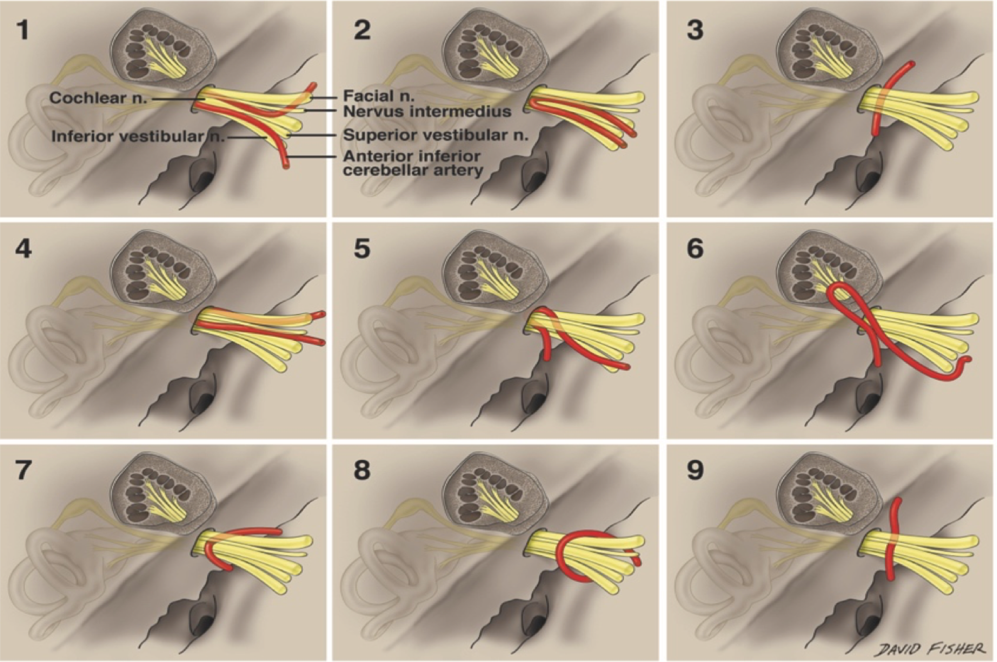

Interestingly, in patients with atresia of the IAC, the facial nerve may take an aberrant course to establish facial motor function.

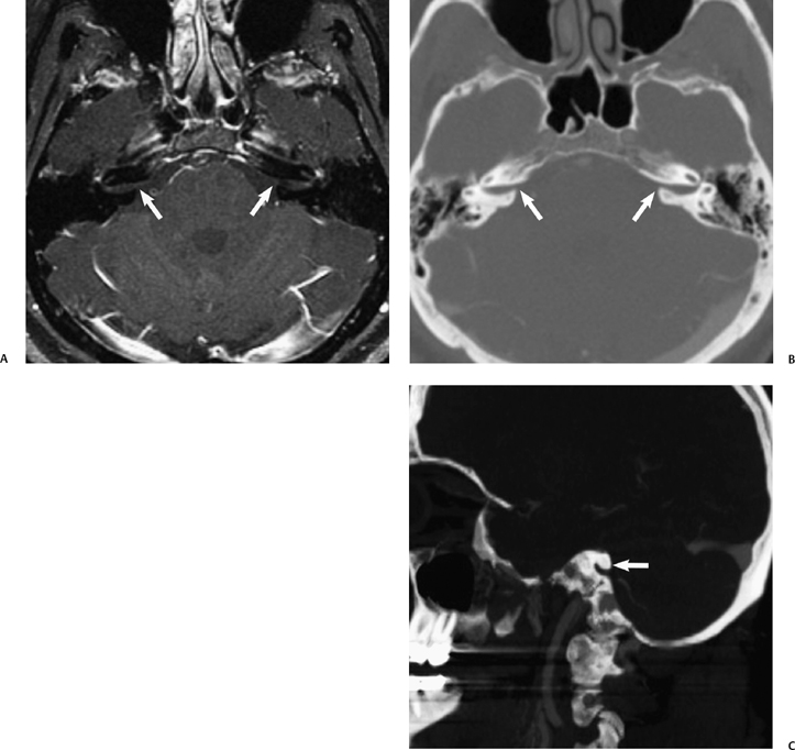
A narrow IAC may accompany inner ear malformations or may be the sole radiographically detectable anomaly in a deaf child. When a patient has normal facial function and an IAC less than 3 mm in diameter, it is likely that the bony canal transmits only the facial nerve ( Fig. Jackler, in Cummings Pediatric Otolaryngology (Second Edition), 2021 Narrow Internal Auditory CanalĪ narrow IAC may indicate a failure of eighth cranial nerve development. The basiocciput is a large quadrilateral portion of the occipital bone that extends anterosuperiorly from the anterior margin of the foramen magnum to reach the sphenoid bone ~ 2/3 of the way up the clivus. In this CT through the condylar fossa of the temporomandibular joint, the petrooccipital fissure is seen between the basiocciput and the temporal bone. Horizontal petrous internal carotid artery canal In this CT through the mid internal auditory canal, the petrooccipital fissure is visible, separating the basioccipital portion of the occipital bone from the temporal bone. The upper carotid space therefore contains CNIX-XII as well as the internal jugular vein. In this CT of the left skull base and temporal bone, notice both the hypoglossal canal and the jugular foramen “empty” into the cephalad carotid space. Again note the lack of definable middle ear structures. The geniculate ganglion is barely visible above and lateral to the cochlea. The area of the tegmen tympani is marked but no landmarks in the middle ear are visible because both air and bone are low signal on MR imaging.Īt the level of the cochlea, the snail shape is particularly obvious displaying both the first and second turns. In this image through the internal auditory canal, an unusually long crista falciformis is seen in the fundus. Notice the superior and lateral semicircular canals adjacent to the vestibule.

The membranous labyrinth of the inner ear is visible as high-signal fluid. This more anterior position persists across the cerebellopontine angle cistern.įirst of 3 coronal T2 MR images of the left ear is shown, presented from posterior to anterior. The CPA cistern segment of the facial nerve is visible in this image just anterior to the vestibulocochlear nerve. It terminates in the geniculate ganglion (anterior genu), then turns and becomes the tympanic segment of the facial nerve which projects posterolaterally to run underneath the lateral semicircular canal.

In this image the labyrinthine segment can be seen passing just superior to the cochlea in the bony labyrinth. At the fundus of the IAC the anterior curving soft tissue structure is the labyrinthine segment of the facial nerve. In this image the internal auditory canal segment of the facial nerve is visible in the anterosuperior quadrant. First of 3 axial high-resolution thin-section T2 MR images of left ear presented from superior to inferior.


 0 kommentar(er)
